Labeled Muscles In The Body Diagram - Pictures: female muscle diagram | Muscle Diagram Female ... - View, isolate, and learn human anatomy structures with zygote body.
Labeled Muscles In The Body Diagram - Pictures: female muscle diagram | Muscle Diagram Female ... - View, isolate, and learn human anatomy structures with zygote body.. For instance, movement about the sagittal axis occurs in the sagittal plane e.g. O skeletal hundreds of randomly located nuclei per cell (cells fuse together in development) striated. Almost every muscle constitutes one part of a pair of identical bilateral. This quiz requires labeling, so it will test your knowledge on how to identify these muscles (latissimus dorsi, trapezius, deltoid, biceps brachii. Now label the diagram in your workbook!
Zygote body is a free online 3d anatomy atlas. Studying these is an ideal first step before moving labeled diagram. Bending forwards at the waist (known as flexion) and. This is what happens in the body. Skeletal, smooth and cardiac, according to the nih.
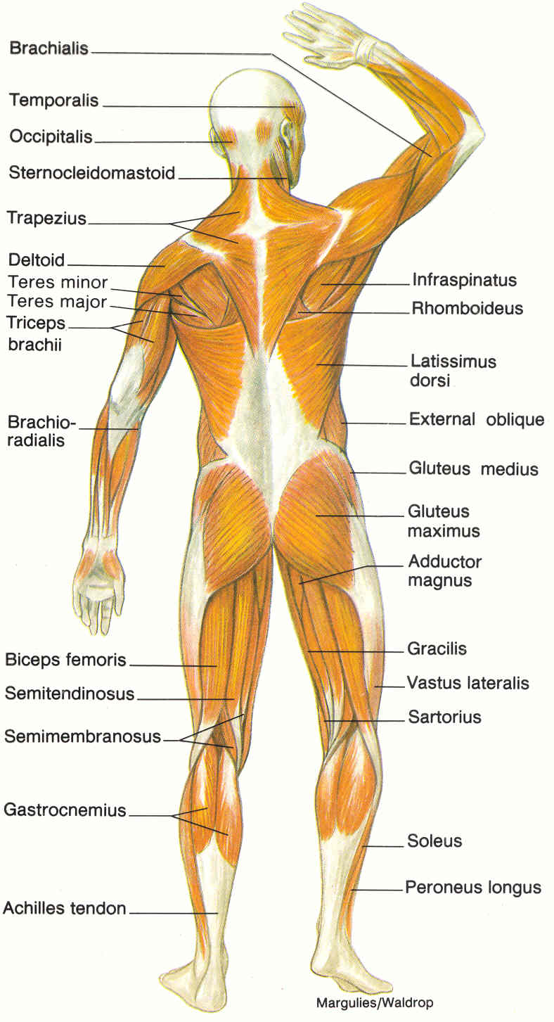
7 best Hand Bones Anatomy images on Pinterest | Hand bone ... from i.pinimg.com This muscular system picture shows all the major muscle groups on the human body from the frontal view. This is an online quiz called label the muscle cell diagram. For instance, movement about the sagittal axis occurs in the sagittal plane e.g. This is what happens in the body. The integumentary system consists of the skin, sweat and oil glands, nails, and hair. View, isolate, and learn human anatomy structures with zygote body. You will also find extensor digitorum, extensor carpi group, latissimus dorsi, external oblique, gluteus medius, gluteus maximus, sartorius, peroneus longus, achilles tendon, gastrocnemius, hamstring group, flexor digitorum, triceps brachii, deltoid, trapezius, sternocleidomastoid, occipitalis in the. O cardiac striated muscle (regular array of actin and myosin in the sarcomeres) each muscle has a single, centered nucleus cells are connected by intercalated discs.
Motion about these planes can be described by an axis of movement.
This is an online quiz called label the muscle cell diagram. We think this is the most useful anatomy picture that you need. Enchantedlearning.com label the body diagram label the human body diagram using the word list below. This quiz focuses on the 23 largest muscles—the ones that account for most of your mobility and strength. Muscle anatomy quiz for anatomy and physiology! Muscles allow us to move and function. Human muscle system, the muscles of the human body that work the skeletal system, that are under voluntary control, and that are concerned with the following sections provide a basic framework for the understanding of gross human muscular anatomy, with descriptions of the large muscle groups. When you are taking anatomy and physiology you will be required to identify major muscles in the human body. Robins are cute, little songbirds in the united states. This quiz requires labeling, so it will test your knowledge on how to identify these muscles (latissimus dorsi, trapezius, deltoid, biceps brachii. Skin is the largest organ in the body and is made up of three layers: View, isolate, and learn human anatomy structures with zygote body. O cardiac striated muscle (regular array of actin and myosin in the sarcomeres) each muscle has a single, centered nucleus cells are connected by intercalated discs.
Motion about these planes can be described by an axis of movement. Muscles allow us to move and function. Studying these is an ideal first step before moving labeled diagram. Rotation and hold ctrl down to pan the view. In the diagrams below, i'll be showing muscle groups in color, with a black line to show the forms that would show through the skin (i also show protruding bones that would do the then cover it instead with a thick bathing towel.
Muscle Diagram | You Can Do More! from youcandomore1.files.wordpress.com This muscle diagram is interactive: O smooth single nuclei not striated. Calcium, iron, and energy in the form of fat. To see a muscular system diagram from the posterior (back) view click here. Anterior muscles in the body. Everyone should identify the location of skeletal muscles in the trunk and upper extremities of the body. We have a lot of muscles in our bodies (literally, over 600). Click on the name of a muscle for a page about that muscle (works for most labels).
Rotation and hold ctrl down to pan the view.
Rotation and hold ctrl down to pan the view. Muscle diagrams are a great way to get an overview of all of the muscles within a body region. We have a lot of muscles in our bodies (literally, over 600). There are over 630 muscles in the human body; This image added by admin. The skeletal system also provides attachment points for muscles to allow movements at the joints. The pelvic floor muscles provide foundational support for the intestines and bladder. O smooth single nuclei not striated. O cardiac striated muscle (regular array of actin and myosin in the sarcomeres) each muscle has a single, centered nucleus cells are connected by intercalated discs. The home button resets the view. This quiz requires labeling, so it will test your knowledge on how to identify these muscles (latissimus dorsi, trapezius, deltoid, biceps brachii. The free muscular system labeling sheet includes a blank diagram to label some of the main muscles in the body. We think this is the most useful anatomy picture that you need.
See how all sharpness disappears? The home button resets the view. Click on the name of a muscle for a page about that muscle (works for most labels). Rotation and hold ctrl down to pan the view. We have a lot of muscles in our bodies (literally, over 600).
diagram of muscular system : Biological Science Picture ... from pulpbits.net Free online quiz label the muscle cell diagram. This is a table of skeletal muscles of the human anatomy. O smooth single nuclei not striated. Learn vocabulary, terms and more with flashcards, games and other study tools. Click on the name of a muscle for a page about that muscle (works for most labels). This is what happens in the body. We think this is the most useful anatomy picture that you need. Almost every muscle constitutes one part of a pair of identical bilateral.
The free muscular system labeling sheet includes a blank diagram to label some of the main muscles in the body.
The soleus connects your lower leg bones to your heel, but it also gives your heart some help by pumping blood back. Anatomynote.com found labelled diagram of the muscles in the human body from plenty of anatomical pictures on the internet. Change from capsule to orbit mode in the upper right to enable full 3d. Muscles allow us to move and function. O cardiac striated muscle (regular array of actin and myosin in the sarcomeres) each muscle has a single, centered nucleus cells are connected by intercalated discs. Label the muscles of the anterior forearm. Everyone should identify the location of skeletal muscles in the trunk and upper extremities of the body. There are over 630 muscles in the human body; Studying these is an ideal first step before moving labeled diagram. This muscular system picture shows all the major muscle groups on the human body from the frontal view. We think this is the most useful anatomy picture that you need. Enchantedlearning.com label the body diagram label the human body diagram using the word list below. Free resources for learning about robins.
The muscles of the shoulder and back chart shows how the many layers of muscle in the shoulder and back are intertwined with the other relevant systems and muscles in adjacent areas like the spine muscles in the body diagram. The free muscular system labeling sheet includes a blank diagram to label some of the main muscles in the body.